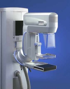
The purpose of a mammogram is to identify abnormalities or changes in breast tissue. Screening mammograms can detect these abnormalities at an early stage resulting in potentially better therapeutic options and possibly less aggressive treatments.
The Mayo Clinic recommends a three-tiered screening mammogram approach:
- Breast health awareness, which includes a woman becoming familiar with her breasts in order to identify breast abnormalities or changes, and to inform her doctor of any changes that may need further evaluation
- Clinical breast exam performed by a health care provider and recommended annually
- Screening mammography beginning at age 40
This approach is consistent with the recommendations of the American Cancer Society. A 2009 study, by the U.S. Prevention Services Task Force that recommended waiting until age 50 to begin screening mammography remains controversial.

X-ray vs. Digital Mammogram Techniques
All mammogram images are captured using X-ray technology. During a traditional mammogram, the results are then viewed on X-ray film. Digital mammograms rely on those same X-ray images, but they are stored on a computer and are analyzed using specialized programs.
Digital mammograms are said to offer better results for:
Women <50 years of age
Women with dense breasts (more tissue, less fat)
Women before or <1 year into the menopause cycle
With either method, you’ll be put through the same steps. Your breasts will be flattened or compressed. While you stand still holding your breath, a technician will take X-ray images. The entire process should last about 15 to 20 minutes.
A woman’s risk of developing breast cancer increases, as she gets older. The risk of breast cancer, however, is not the same for all women in a given age group. Research has shown that women with the following risk factors have an increased chance of developing breast cancer:
- Advancing age – most important risk factor
- Personal history of breast cancer
- Family history of breast cancer
- Radiation exposure to the chest
- Obesity
- Starting menstruation before age 12
- Beginning menopause after age 55
- Having your first child after age 35
However, a majority of women who develop breast cancer have no family history or risk factors.
If you’re concerned about screening mammograms, talk to Dr. Forley or your primary care doctor and learn what’s right for you based on your individual risks. It’s important that the two of you work together to develop a screening plan.
Tags: Breast Augmentation, breast cancer, breast implants
Written by Dr. Forley on June 25, 2012


